





Expo
view channel
view channel
view channel
view channel
view channel
view channel
view channel
view channel
AICritical CareSurgical TechniquesPatient Care
Health ITPoint of CareBusiness
Events
Webinars

- 3D-Printed Membrane Could Enable Smaller, More Efficient Artificial Lungs
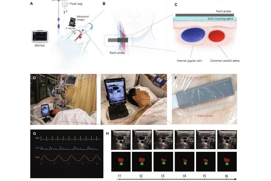
- Wearable Ultrasound Patch Uses AI to Detect Elevated Central Venous Pressure
- Switchable Soft Material Could Improve On-Demand Drug Delivery
- Smart Patch Could Detect Fentanyl and Deliver Automatic Overdose Aid
- AI Wearable Enables Continuous Noninvasive Arterial Blood Pressure Monitoring
- Heartbeat-Powered Pacemaker Could Eliminate Battery Replacement Surgery
- 3D Bioprinting Method Enhances Bone Repair by Promoting Blood Vessel Growth
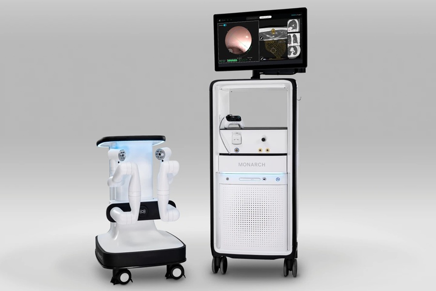
- New Robotic Bronchoscopy Software Enhances Lung Nodule Navigation and Targeting
- New Radiofrequency Wand Enhances Access and Tissue Targeting in Hip Surgery
- Hybrid Bioprinting Platform Creates Capillary-Scale Vascular Networks
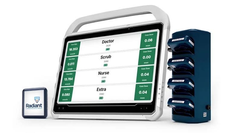
- Real-Time Radiation Monitor Supports Safer Fluoroscopy-Guided Procedures
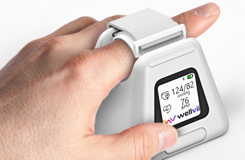
- FDA-Cleared Finger Cuff Device Enables Home Blood Pressure Monitoring
- AI Avatar Doctor Improves Patient Understanding Before Radiotherapy
- Wearable Sleep Data Predict Adherence to Pulmonary Rehabilitation
- Revolutionary Automatic IV-Line Flushing Device to Enhance Infusion Care
- Imaging Precision Tool Enables Method Comparison Without Ground-Truth Data
- AI-Derived Brain Age from MRI Predicts Future Dementia Risk
- Imaging Biomarkers Aim to Personalize Treatment for Post-Stroke Bladder Dysfunction
- New AI Method Improves CT Images by Reducing Metal Artifacts
- MRI-Guided Ultrasound Method Advances Targeted Treatment of Gliomas
- MIMEDX to Add Sanara MedTech’s Regenerative Technologies Through $350 Million Deal
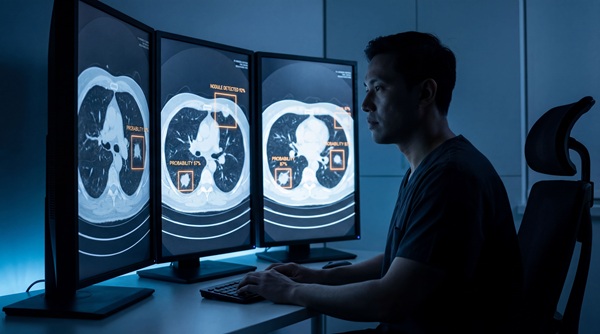
- AI Software Combines Nodule Detection and Diagnosis for Lung Cancer Screening
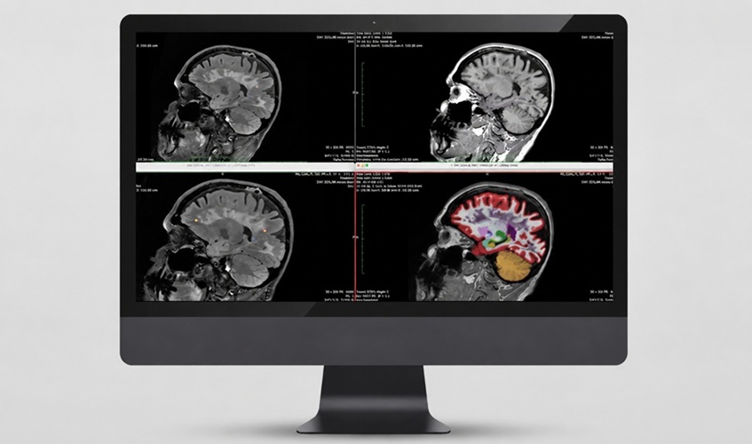
- AI Care Platform Adds Quantitative MRI Analysis for Multiple Sclerosis
- Danaher Completes Acquisition of Masimo to Expand Patient Monitoring Capabilities
- Endologix Adds FDA-Cleared Peripheral Thrombectomy System
- Digital Symptom Monitoring Supports More Personalized Breast Cancer Care
- Short Digital Training Program Improves Motor Function in Parkinson’s Disease
- New Cloud Platform Streamlines Capsule Endoscopy Workflow and Reporting
- Smartwatch Monitoring Identifies Abnormal Vital Signs Before Routine Checks
- Weekly Remote Symptom Monitoring Improves Symptom Control in Advanced Cancer
- AI Digital Twin Platform Supports Personalized Cancer Treatment
- Study Reveals Persistent Race and Gender Bias in Medical AI Models
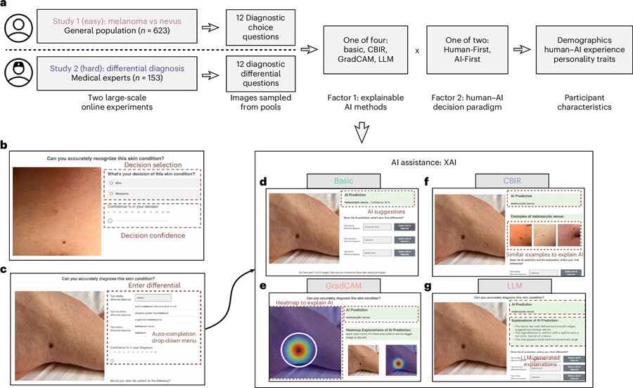
- New Study Highlights Benefits and Risks of AI Explanations in Clinical Diagnosis
- AI Improves Bronchoscopic Navigation to Difficult-to-Reach Lung Lesions
- Collaborative AI System Improves Medical Diagnostic Accuracy
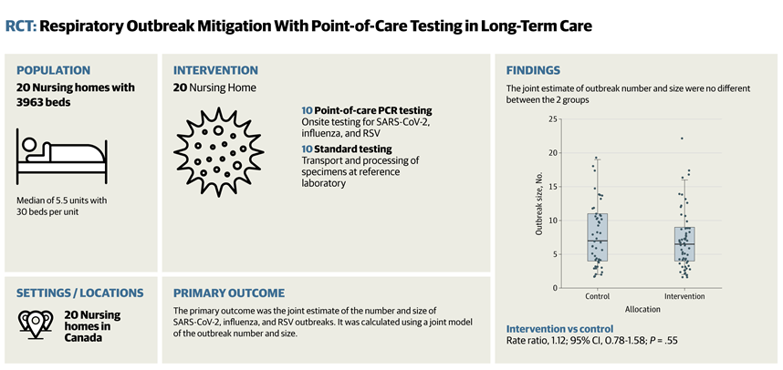
- Rapid Onsite Testing Reduces Emergency Department Transfers in Long-Term Care
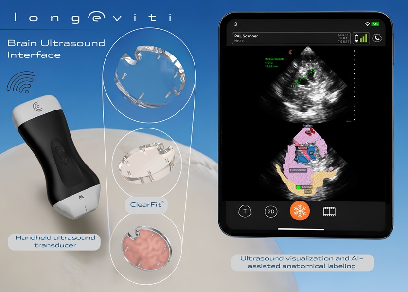
- New Brain Ultrasound Platform Enables Bedside Postoperative Imaging
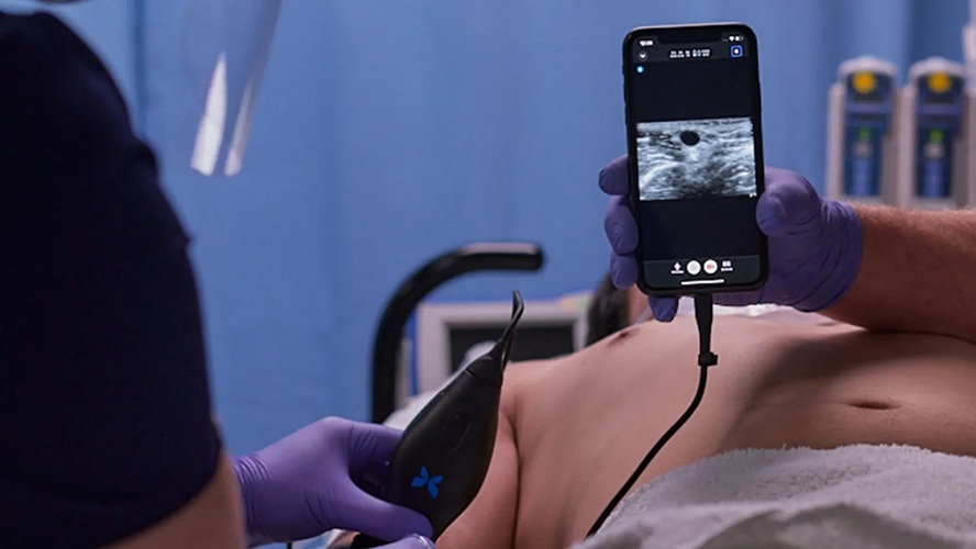
- Handheld Ultrasound Expands Point-of-Care Imaging Access in Brazil
- AI Dermatology Platform Targets Early Detection of Non-Melanoma Skin Cancer
- Handheld AI Device for Point-of-Care Skin Lesion Assessment Receives CE Mark
 Expo
Expo
- 3D-Printed Membrane Could Enable Smaller, More Efficient Artificial Lungs
- Wearable Ultrasound Patch Uses AI to Detect Elevated Central Venous Pressure
- Switchable Soft Material Could Improve On-Demand Drug Delivery
- Smart Patch Could Detect Fentanyl and Deliver Automatic Overdose Aid
- AI Wearable Enables Continuous Noninvasive Arterial Blood Pressure Monitoring
- Heartbeat-Powered Pacemaker Could Eliminate Battery Replacement Surgery
- 3D Bioprinting Method Enhances Bone Repair by Promoting Blood Vessel Growth
- New Robotic Bronchoscopy Software Enhances Lung Nodule Navigation and Targeting
- New Radiofrequency Wand Enhances Access and Tissue Targeting in Hip Surgery
- Hybrid Bioprinting Platform Creates Capillary-Scale Vascular Networks
- Real-Time Radiation Monitor Supports Safer Fluoroscopy-Guided Procedures
- FDA-Cleared Finger Cuff Device Enables Home Blood Pressure Monitoring
- AI Avatar Doctor Improves Patient Understanding Before Radiotherapy
- Wearable Sleep Data Predict Adherence to Pulmonary Rehabilitation
- Revolutionary Automatic IV-Line Flushing Device to Enhance Infusion Care
- Imaging Precision Tool Enables Method Comparison Without Ground-Truth Data
- AI-Derived Brain Age from MRI Predicts Future Dementia Risk
- Imaging Biomarkers Aim to Personalize Treatment for Post-Stroke Bladder Dysfunction
- New AI Method Improves CT Images by Reducing Metal Artifacts
- MRI-Guided Ultrasound Method Advances Targeted Treatment of Gliomas
- MIMEDX to Add Sanara MedTech’s Regenerative Technologies Through $350 Million Deal
- AI Software Combines Nodule Detection and Diagnosis for Lung Cancer Screening
- AI Care Platform Adds Quantitative MRI Analysis for Multiple Sclerosis
- Danaher Completes Acquisition of Masimo to Expand Patient Monitoring Capabilities
- Endologix Adds FDA-Cleared Peripheral Thrombectomy System
- Digital Symptom Monitoring Supports More Personalized Breast Cancer Care
- Short Digital Training Program Improves Motor Function in Parkinson’s Disease
- New Cloud Platform Streamlines Capsule Endoscopy Workflow and Reporting
- Smartwatch Monitoring Identifies Abnormal Vital Signs Before Routine Checks
- Weekly Remote Symptom Monitoring Improves Symptom Control in Advanced Cancer
- AI Digital Twin Platform Supports Personalized Cancer Treatment
- Study Reveals Persistent Race and Gender Bias in Medical AI Models
- New Study Highlights Benefits and Risks of AI Explanations in Clinical Diagnosis
- AI Improves Bronchoscopic Navigation to Difficult-to-Reach Lung Lesions
- Collaborative AI System Improves Medical Diagnostic Accuracy
- Rapid Onsite Testing Reduces Emergency Department Transfers in Long-Term Care
- New Brain Ultrasound Platform Enables Bedside Postoperative Imaging
- Handheld Ultrasound Expands Point-of-Care Imaging Access in Brazil
- AI Dermatology Platform Targets Early Detection of Non-Melanoma Skin Cancer
- Handheld AI Device for Point-of-Care Skin Lesion Assessment Receives CE Mark



























 Client Login