Expo
view channel
view channel
view channel
view channel
view channel
Medical Imaging
AICritical CareSurgical TechniquesPatient Care
Point of CareBusiness
Events

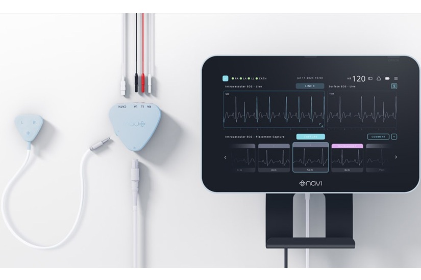
- World-First Technology Uses Real-Time ECG Signal Analysis for Accurate CVAD Placement
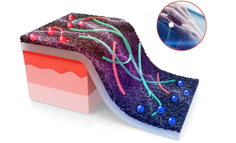
- Smart Sensor Enables Precise, Self-Powered Tracking of Healing Wounds
- AI Outperforms Humans at Analyzing Long-Term ECG Recordings
- Skin Patch Activates New Gene Switch to Treat Diabetes
- Zinc-Based Dissolvable Implants to Transform Bone Repair
- "Ultra-Rapid" Testing in the OR Could Enable Accurate Removal of Brain Tumors
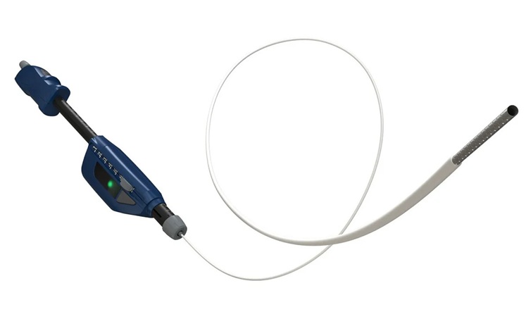
- Automated Endoscopic Device Obtains Improved Biopsy Results in Single Pass
- World's First Machine Learning Model Combats Wrong-Site Surgery
- Novel Method Combining Heart Biopsy and Device Implantation Reduces Complications Risk
- New Surface Coating Could Prevent Blood Clotting in Medical Devices and Implants
- First-Of-Its-Kind Portable Germicidal Light Technology Disinfects High-Touch Clinical Surfaces in Seconds
- Surgical Capacity Optimization Solution Helps Hospitals Boost OR Utilization
- Game-Changing Innovation in Surgical Instrument Sterilization Significantly Improves OR Throughput
- Next Gen ICU Bed to Help Address Complex Critical Care Needs
- Groundbreaking AI-Powered UV-C Disinfection Technology Redefines Infection Control Landscape
- Teleflex to Acquire BIOTRONIK’s Vascular Intervention Business
- Philips and Mass General Brigham Collaborate on Improving Patient Care with Live AI-Powered Insights
- Arab Health 2025 Celebrates Landmark 50th Edition
- Boston Scientific Acquires Medical Device Company Intera Oncology
- MEDICA 2024 to Highlight Hot Topics of MedTech Industry
- Smartwatches Could Detect Congestive Heart Failure
- Versatile Smart Patch Combines Health Monitoring and Drug Delivery
- Machine Learning Model Improves Mortality Risk Prediction for Cardiac Surgery Patients
- Strategic Collaboration to Develop and Integrate Generative AI into Healthcare
- AI-Enabled Operating Rooms Solution Helps Hospitals Maximize Utilization and Unlock Capacity

Expo
 view channel
view channel
view channel
view channel
Medical Imaging
AICritical CareSurgical TechniquesPatient Care
Point of CareBusiness
Events
Advertise with Us
view channel
view channel
view channel
view channel
view channel
Medical Imaging
AICritical CareSurgical TechniquesPatient Care
Point of CareBusiness
Events
Advertise with Us


- World-First Technology Uses Real-Time ECG Signal Analysis for Accurate CVAD Placement
- Smart Sensor Enables Precise, Self-Powered Tracking of Healing Wounds
- AI Outperforms Humans at Analyzing Long-Term ECG Recordings
- Skin Patch Activates New Gene Switch to Treat Diabetes
- Zinc-Based Dissolvable Implants to Transform Bone Repair
- "Ultra-Rapid" Testing in the OR Could Enable Accurate Removal of Brain Tumors
- Automated Endoscopic Device Obtains Improved Biopsy Results in Single Pass
- World's First Machine Learning Model Combats Wrong-Site Surgery
- Novel Method Combining Heart Biopsy and Device Implantation Reduces Complications Risk
- New Surface Coating Could Prevent Blood Clotting in Medical Devices and Implants
- First-Of-Its-Kind Portable Germicidal Light Technology Disinfects High-Touch Clinical Surfaces in Seconds
- Surgical Capacity Optimization Solution Helps Hospitals Boost OR Utilization
- Game-Changing Innovation in Surgical Instrument Sterilization Significantly Improves OR Throughput
- Next Gen ICU Bed to Help Address Complex Critical Care Needs
- Groundbreaking AI-Powered UV-C Disinfection Technology Redefines Infection Control Landscape
- Teleflex to Acquire BIOTRONIK’s Vascular Intervention Business
- Philips and Mass General Brigham Collaborate on Improving Patient Care with Live AI-Powered Insights
- Arab Health 2025 Celebrates Landmark 50th Edition
- Boston Scientific Acquires Medical Device Company Intera Oncology
- MEDICA 2024 to Highlight Hot Topics of MedTech Industry
- Smartwatches Could Detect Congestive Heart Failure
- Versatile Smart Patch Combines Health Monitoring and Drug Delivery
- Machine Learning Model Improves Mortality Risk Prediction for Cardiac Surgery Patients
- Strategic Collaboration to Develop and Integrate Generative AI into Healthcare
- AI-Enabled Operating Rooms Solution Helps Hospitals Maximize Utilization and Unlock Capacity


