Expo
view channel
view channel
view channel
view channel
Medical Imaging
AICritical Care
Patient CareHealth ITPoint of CareBusiness
Events
Webinars

- Brain-Based Biomarker Could Predict Alzheimer’s Disease Progression
- AI Model Detects Hidden Diabetes Risk by Reading Glucose Spikes
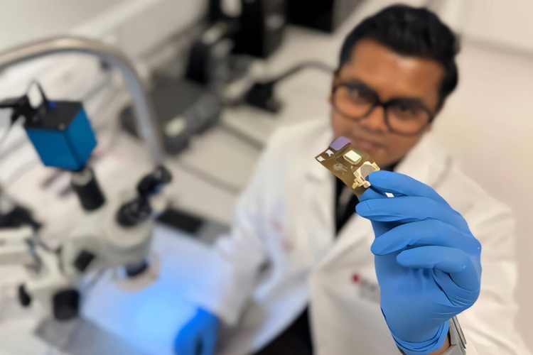
- Wearable Wound Monitoring Device to Improve Chronic Infection Care
- World's First Wearable-Powered System Predicts Acute Inflammation With 90% Sensitivity
- Swallowable Biosensor Pill Tracks Inflammatory Bowel Disease
- AI Cuts Diagnostic Delays in Prostate Cancer
- Surgical Micro-Robot Sees and Corrects Movements from Within
- 'Google Maps' for Surgeons to Help Perform Complex Robot-Assisted Esophagectomy
- New Technique for Measuring Brain Blood Flow During Surgery Can Prevent Strokes
- Computational Model Accurately Simulates Shunt Performance to Prevent Repeat Surgeries
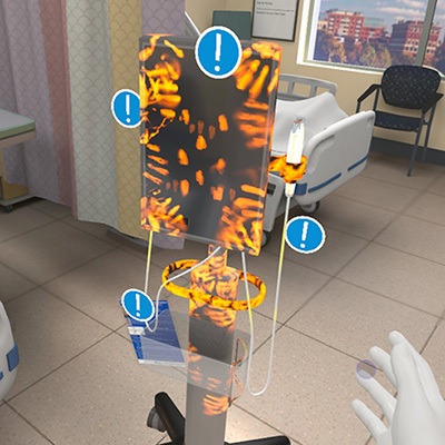
- VR Training Tool Combats Contamination of Portable Medical Equipment
- Portable Biosensor Platform to Reduce Hospital-Acquired Infections
- First-Of-Its-Kind Portable Germicidal Light Technology Disinfects High-Touch Clinical Surfaces in Seconds
- Surgical Capacity Optimization Solution Helps Hospitals Boost OR Utilization
- Game-Changing Innovation in Surgical Instrument Sterilization Significantly Improves OR Throughput
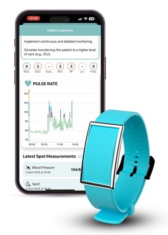
- Medtronic Partners with Corsano to Expand Acute Care & Monitoring Portfolio in Europe
- Expanded Collaboration to Transform OR Technology Through AI and Automation
- Becton Dickinson to Spin Out Biosciences and Diagnostic Solutions Business
- Boston Scientific Acquires Medical Device Company SoniVie
- 2026 World Hospital Congress to be Held in Seoul

 Expo
Expo
- Brain-Based Biomarker Could Predict Alzheimer’s Disease Progression
- AI Model Detects Hidden Diabetes Risk by Reading Glucose Spikes
- Wearable Wound Monitoring Device to Improve Chronic Infection Care
- World's First Wearable-Powered System Predicts Acute Inflammation With 90% Sensitivity
- Swallowable Biosensor Pill Tracks Inflammatory Bowel Disease
- AI Cuts Diagnostic Delays in Prostate Cancer
- Surgical Micro-Robot Sees and Corrects Movements from Within
- 'Google Maps' for Surgeons to Help Perform Complex Robot-Assisted Esophagectomy
- New Technique for Measuring Brain Blood Flow During Surgery Can Prevent Strokes
- Computational Model Accurately Simulates Shunt Performance to Prevent Repeat Surgeries
- VR Training Tool Combats Contamination of Portable Medical Equipment
- Portable Biosensor Platform to Reduce Hospital-Acquired Infections
- First-Of-Its-Kind Portable Germicidal Light Technology Disinfects High-Touch Clinical Surfaces in Seconds
- Surgical Capacity Optimization Solution Helps Hospitals Boost OR Utilization
- Game-Changing Innovation in Surgical Instrument Sterilization Significantly Improves OR Throughput
- Medtronic Partners with Corsano to Expand Acute Care & Monitoring Portfolio in Europe
- Expanded Collaboration to Transform OR Technology Through AI and Automation
- Becton Dickinson to Spin Out Biosciences and Diagnostic Solutions Business
- Boston Scientific Acquires Medical Device Company SoniVie
- 2026 World Hospital Congress to be Held in Seoul


